Scientists grow 5-week-old human brain

Scientists successfully grow human brain in lab

Scientists successfully grow human brain in lab

Growing brain tissue in a dish has been done before, but bold new research announced this week shows that scientists’ ability to create human brains in laboratory settings has come a long way quickly.

Researchers at the Ohio State University in the US claim to have developed the most complete laboratory-grown human brain ever, creating a model with the brain maturity of a 5-week-old foetus. The brain, which is approximately the size of a pencil eraser, contains 99 percent of the genes that would be present in a natural human foetal brain.

“It not only looks like the developing brain, its diverse cell types express nearly all genes like a brain,” Rene Anand, professor of biological chemistry and pharmacology at Ohio State and lead researcher on the brain model, said in a statement.

“We’ve struggled for a long time trying to solve complex brain disease problems that cause tremendous pain and suffering. The power of this brain model bodes very well for human health because it gives us better and more relevant options to test and develop therapeutics other than rodents.”

Anand turned to stem cell engineering four years ago after his specialized field of research – examining the relationship between nicotinic receptors and central nervous system disorders – ran into complications using rodent specimens. Despite having limited funds, Anand and his colleagues succeeded with their proprietary technique, which they are in the process of commercializing.

The brain they have developed is a virtually complete recreation of a human foetal brain, primarily missing only a vascular system – in other words, all the blood vessels. But everything else (spinal cord, major brain regions, multiple cell types, signalling circuitry is there). What’s more, it’s functioning, with high-resolution imaging of the brain model showing functioning neurons and brain cells.

The researchers say that it takes 15 weeks to grow a lab-developed brain to the equivalent of a 5-week-old foetal human brain, and the longer the maturation process the more complete the organoid will become.

“If we let it go to 16 or 20 weeks, that might complete it, filling in that 1 percent of missing genes. We don’t know yet,” said Anand.

The scientific benefit of growing human brains in laboratory settings is that it enables high-end research into human diseases that cannot be completed using rodents.

“In central nervous system diseases, this will enable studies of either underlying genetic susceptibility or purely environmental influences, or a combination,” said Anand. “Genomic science infers there are up to 600 genes that give rise to autism, but we are stuck there. Mathematical correlations and statistical methods are insufficient to in themselves identify causation. You need an experimental system – you need a human brain.”

The research was presented this week at the Military Health System Research Symposium.


Source:  sciencealert.com

Human cartilage grown in lab

Engineers grow functional human cartilage in lab:

Engineers grow functional human cartilage in lab

Engineers grow functional human cartilage in lab


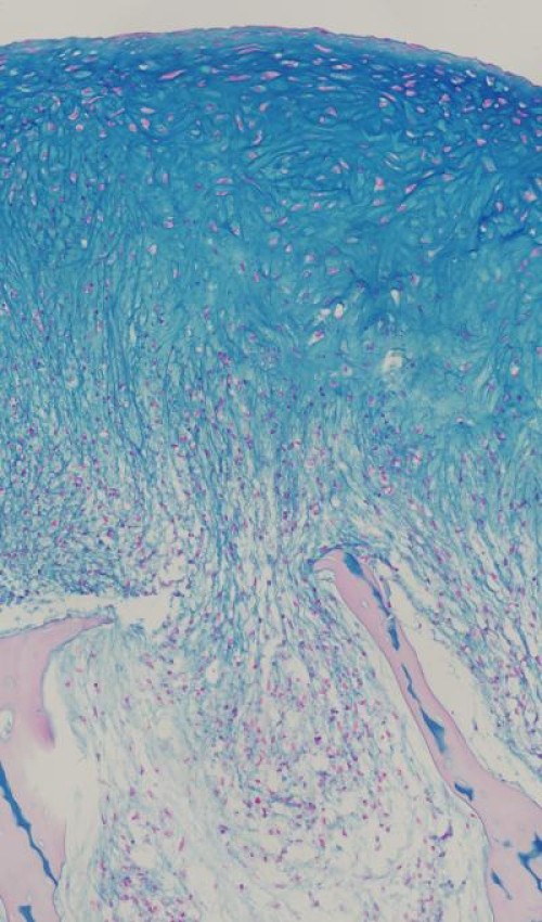
Researchers at Columbia Engineering announced that they have successfully grown fully functional human cartilage in vitro from human stem cells derived from bone marrow tissue. Their study, which demonstrates new ways to better mimic the enormous complexity of tissue development, regeneration, and disease.
“We’ve been able — for the first time — to generate fully functional human cartilage from mesenchymal stem cells by mimicking in vitro the developmental process of mesenchymal condensation,” says Gordana Vunjak-Novakovic, who led the study and is the Mikati Foundation Professor of Biomedical Engineering at Columbia Engineering and professor of medical sciences. “This could have clinical impact, as this cartilage can be used to repair a cartilage defect, or in combination with bone in a composite graft grown in lab for more complex tissue reconstruction.”


For more than 20 years, researchers have unofficially called cartilage the “official tissue of tissue engineering,” Vunjak-Novakovic observes. Many groups studied cartilage as an apparently simple tissue: one single cell type, no blood vessels or nerves, a tissue built for bearing loads while protecting bone ends in the joints. While there has been great success in engineering pieces of cartilage using young animal cells, no one has, until now, been able to reproduce these results using adult human stem cells from bone marrow or fat, the most practical stem cell source.
Vunjak-Novakovic’s team succeeded in growing cartilage with physiologic architecture and strength by radically changing the tissue-engineering approach.
The general approach to cartilage tissue engineering has been to place cells into a hydrogel and culture them in the presence of nutrients and growth factors and sometimes also mechanical loading. But using this technique with adult human stem cells has invariably produced mechanically weak cartilage. So Vunjak-Novakovic and her team, who have had a longstanding interest in skeletal tissue engineering, wondered if a method resembling the normal development of the skeleton could lead to a higher quality of cartilage.


Sarindr Bhumiratana, postdoctoral fellow in Vunjak-Novakovic’s Laboratory for Stem Cells and Tissue Engineering, came up with a new approach: inducing the mesenchymal stem cells to undergo a condensation stage as they do in the body before starting to make cartilage. He discovered that this simple but major departure from how things were usually? being done resulted in a quality of human cartilage not seen before.


Gerard Ateshian, Andrew Walz Professor of Mechanical Engineering, professor of biomedical engineering, and chair of the Department of Mechanical Engineering, and his PhD student, Sevan Oungoulian, helped perform measurements showing that the lubricative property and compressive strength — the two important functional properties — of the tissue-engineered cartilage approached those of native cartilage.
The researchers then used their method to regenerate large pieces of anatomically shaped and mechanically strong cartilage over the bone, and to repair defects in cartilage.


“Our whole approach to tissue engineering is biomimetic in nature, which means that our engineering designs are defined by biological principles,” Vunjak-Novakovic notes. “This approach has been effective in improving the quality of many engineered tissues — from bone to heart. Still, we were really surprised to see that our cartilage, grown by mimicking some aspects of biological development, was as strong as ‘normal’ human cartilage.”


The team plans next to test whether the engineered cartilage tissue maintains its structure and long-term function when implanted into a defect.


“This is a very exciting time for tissue engineers,” says Vunjak-Novakovic. “Stem cells are transforming the future of medicine, offering ways to overcome some of the human body’s fundamental limitations. We bioengineers are now working with stem cell scientists and clinicians to develop technologies that will make this dream possible. This project is a wonderful example that we need to ‘think as a cell’ to find out how exactly to coax the cells into making a functional human tissue of a specific kind. It’s emblematic of the progress being driven by the exceptional young talent we have among our postdocs and students at Columbia Engineering.”


Source:  sciencedaily.com

Scientist’s Grow Liquid Crystal

Researchers Grow Liquid Crystal:

Researchers Grow Liquid Crystal

Researchers Grow Liquid Crystal


In previous studies , the team produced patterns “defects” votes disruptions repeating patterns found in liquid crystals, in grids and rings nanometer scale. A three-dimensional matrix in the form of a flower : The new study a more complex pattern of an even simpler template is added.

And because the petals of this ” bloom ” are made transparent liquid crystal and radiates outward in a circle from a center point , the assembly resembles a compound eye and therefore can be used as a lens .

The team consists of Randall Kamien , a professor in the School of Arts and the Department of Physical Sciences and Astronomy ; Kathleen Stebe , the School of Engineering and Associate Dean of Applied Sciences for research and professor of Chemical and Bio molecular Engineering and Shu Yang , professor of Engineering departments Materials Science and Engineering and Chemical and Bio molecular Engineering . Members of their laboratories also contributed to the new study, including lead author Daniel Beller , Mohamed Gharbi and Apiradee Honglawan .

Ongoing work of researchers with liquid crystals is an example of a growing field of nanotechnology known as “directed assembly” , in which scientists and engineers aim to manufacture structures on smaller scales without that each component individually manipulated . Rather, the starting conditions are set precisely defined and allow the physics and chemistry that govern these components do the rest.

The starting conditions in the experiments of previous investigators were templates consist of small stalls . In one of his studies, which showed that changing the size , shape and spacing of these posts would result in corresponding changes in the patterns of defects on the surface of the liquid crystal resting on top of them . In another experiment , the researchers showed that they could make a ” hula hoop ” defects around the individual poles , which then act as a second model for a ring of surface defects .In his latest work , the researchers used a much simpler signal.

“Before these liquid crystals were growing into something like a trellis, a template with precisely ordered features,” Kamien said. ” Here , we are planting a seed. ”

The seeds , in this case , silica beads were – essentially polished sand grains . Planted on top of a pool of crystal flower-like patterns of liquid defects grow around each bead . The key difference between the template in this experiment and those in the earlier work of the research team was the shape of the interface between the template and the liquid crystal.

In their experiment that generated defects grid patterns , these patterns are derived from the signals generated by microposts templates . Domains elastic energy originated in the tops and edges of these flat positions and traveled to the liquid crystal layer , culminating in defects. Using a cord instead of a message , as the researchers did in their last experiment , made ​​so that the interface is no longer flat .

“Not just the interface at an angle is an angle that keeps changing , ” Kamien said. ” The way in which the liquid crystal responds to it is that makes these petal shapes in smaller sizes and smaller, trying to match the angle of the pearl until everything is flat. ”

The surface tension in the bead also makes it so that these petals are arranged in one of the convexly levels. And because the liquid crystal can interact with light , the whole can function as a lens, focusing the light to a point below the bead.

“It’s like the compound eye of an insect, or mirrors in larger telescopes,” Kamien said. ” As we learn more about these systems , we will be able to do this type of lenses to order and use them to direct the light . ”

This type of directed assembly could be useful in the manufacture of optical switches and other applications .

Scientists Grow Human Brain

Scientists Grow Human Brain From Stem Cells:

Scientists Grow Human Brain From Stem Cells

Scientists Grow Human Brain From Stem Cells

Ear, eye, liver, windpipe, bladder and even a heart. The list of body parts grown from stem cells is getting longer and longer. Now add to it one of the most complex organs: the brain.

A team of European scientists has grown parts of a human brain in tissue culture from stem cells. Their work could help scientists understand the origins of schizophrenia or autism and lead to drugs to treat them, said Juergen Knoblich, deputy scientific director at the Institute of Molecular Biotechnology of the Austrian Academy of Sciences and one of the paper’s co-authors.

The advance could also eliminate the need for conducting experiments on animals, whose brains are not a perfect model for humans.

To grow the brain structures, called organoids, the scientists used stem cells, which can develop into any other kind of cell in the body. They put the stem cells into a special solution designed to promote the growth of neural cells. Bits of gel interspersed throughout the solution gave the cells a three-dimensional structure to grow upon. In eight to 10 days, the stem cells turned into brain cells. After 20 days to a month, the cells matured into a size between three and four millimeters, representing specific brain regions such as the cortex and the hindbrain.

Growing brain tissue this way marks a major advancement because the lab-grown brain cells self-organized and took on growth patterns seen in a developing, fetal brain.

Currently, the organoids are limited on how big they can get because they do not have a circulatory system to move around nutrients.

Knoblich’s team didn’t stop at growing the brain organoids, though. They went a step further and used the developing tissue to study microcephaly, a condition in which the brain stops growing. Microcephalic patients are born with smaller brains and impaired cognitive development. Studying microcephaly in mice doesn’t help because human and mouse brains are too different.

For this part of the study, the researchers used stem cells from a microcephalic patient and grew neurons in a culture. They found that normal brains have progenitor stem cells that make neurons and can do so repeatedly. In microcephalic brains, the progenitor cells differentiate into neurons earlier, said Madeline A. Lancaster, the study’s lead author. The brain doesn’t make as many neurons and a child is born with a much smaller brain volume.

Yoshiki Sasai, a stem-cell biologist at the Riken Center for Developmental Biology in Kobe, Japan, garnered headlines last year by growing the precursors to a human eye.

“The most important advancement is that they combined this self-organization culture with disease-specific cells to model a genetic disease of human brain malformation,” he said.

“Everything we have done with other organs starts with this stage,” said Dr. Anthony Atala, the director of the Wake Forest Institute for Regenerative Medicine, who has done years of research on using 3D printers to build organs. Atala was not involved in this study, but he noted that before he could build organs, he needed to grow the pieces in order to get the cells to differentiate in just the right way. So though it’s unlikely anyone will print brains the way he did a kidney, this kind of experiment is where organ regeneration starts.

Knoblich said the next step is studying other brain disorders, but it will take some time to grow enough brain tissue. One factor is maximum size and how far the brain can develop in the culture. Brain cells develop in layers, and there are several by the time a baby is born. The cortical cells Knoblich’s team grew only had one such layer. Another factor is getting blood vessels inside the tissue. That problem could be solved some time in the future, though he said he couldn’t predict when.

It is tempting to think one day there will be whole brains in vats, but that isn’t likely to happen.

“Aside from the severe ethical problem, I do not think this will be possible,” Knoblich said. To form actual functioning neural circuits, a brain needs sensory input. “Without any sensory input, the proper organization may not happen.”

GM corn grows horrifying tumors, 70% of females die early

Shock findings in new GMO study: Rats fed lifetime of GM corn grow horrifying tumors, 70% of females die early:

Shock findings in new GMO study: Rats fed lifetime of GM corn grow horrifying tumors, 70% of females die early

Shock findings in new GMO study: Rats fed lifetime of GM corn grow horrifying tumors, 70% of females die early


Eating genetically modified corn (GM corn) and consuming trace levels of Monsanto’s Roundup chemical fertilizer caused rats to develop horrifying tumors, widespread organ damage, and premature death. That’s the conclusion of a shocking new study that looked at the long-term effects of consuming Monsanto’s genetically modified corn. The study has been deemed “the most thorough research ever published into the health effects of GM food crops and the herbicide Roundup on rats.” News of the horrifying findings is spreading like wildfire across the internet, with even the mainstream media seemingly in shock over the photos of rats with multiple grotesque tumors… tumors so large the rats even had difficulty breathing in some cases. GMOs may be the new thalidomide. “Monsanto Roundup weedkiller and GM maize implicated in ‘shocking’ new cancer study” wrote The Grocery, a popular UK publication.  It reported, “Scientists found that rats exposed to even the smallest amounts, developed mammary tumors and severe liver and kidney damage as early as four months in males, and seven months for females.” The Daily Mail reported, “Fresh row over GM foods as French study claims rats fed the controversial crops suffered tumors.”  It goes on to say: “The animals on the GM diet suffered mammary tumors, as well as severe liver and kidney damage. The researchers said 50 percent of males and 70 percent of females died prematurely, compared with only 30 percent and 20 percent in the control group.”

The study, led by Gilles-Eric Seralini of the University of Caen, was the first ever study to examine the long-term (lifetime) effects of eating GMOs. You may find yourself thinking it is absolutely astonishing that no such studies were ever conducted before GM corn was approved for widespread use by the USDA and FDA, but such is the power of corporate lobbying and corporate greed.

Findings from the study

Here are some of the shocking findings from the study:

• Up to 50% of males and 70% of females suffered premature death.

• Rats that drank trace amounts of Roundup (at levels legally allowed in the water supply) had a 200% to 300% increase in large tumors.

• Rats fed GM corn and traces of Roundup suffered severe organ damage including liver damage and kidney damage.

• The study fed these rats NK603, the Monsanto variety of GM corn that’s grown across North America and widely fed to animals and humans. This is the same corn that’s in your corn-based breakfast cereal, corn tortillas and corn snack chips.

The Daily Mail is reporting on some of the reaction to the findings:

France’s Jose Bove, vice-chairman of the European Parliament’s commission for agriculture and known as a fierce opponent of GM, called for an immediate suspension of all EU cultivation and import authorisations of GM crops. ‘This study finally shows we are right and that it is urgent to quickly review all GMO evaluation processes,’ he said in a statement. ‘National and European food security agencies must carry out new studies financed by public funding to guarantee healthy food for European consumers.’

The study is entitled, “A Comparison of the Effects of Three GM Corn Varieties on Mammalian Health.”

That abstract include this text. Note: “hepatorenal toxicity” means toxic to the liver.

Our analysis clearly reveals for the 3 GMOs new side effects linked with GM maize consumption, which were sex- and often dose-dependent. Effects were mostly associated with the kidney and liver, the dietary detoxifying organs, although different between the 3 GMOs. Other effects were also noticed in the heart, adrenal glands, spleen and haematopoietic system. We conclude that these data highlight signs of hepatorenal toxicity, possibly due to the new pesticides specific to each GM corn. In addition, unintended direct or indirect metabolic consequences of the genetic modification cannot be excluded.

Here are some quotes from the researchers:

“This research shows an extraordinary number of tumors developing earlier and more aggressively – particularly in female animals. I am shocked by the extreme negative health impacts.” – Dr Michael Antoniou, molecular biologist, King’s College London.

“We can expect that the consumption of GM maize and the herbicide Roundup, impacts seriously on human health.” – Dr Antoniou.

“This is the first time that a long-term animal feeding trial has examined the impact of feeding GM corn or the herbicide Roundup, or a combination of both and the results are extremely serious. In the male rats, there was liver and kidney disorders, including tumors and even more worryingly, in the female rats, there were mammary tumors at a level which is extremely concerning; up to 80 percent of the female rats had mammary tumors by the end of the trial.” – Patrick Holden, Director, Sustainable Food Trust.